
Dr Christian Engelbrecht
Oogchirurg | Ophthalmic Surgeon

Get to know your surgeon

Dr Christian Engelbrecht grew up in the small town of Leandra in Mpumalanga. He later moved to Pretoria and matriculated from Pretoria Boys High School. Dr Engelbrecht completed his undergraduate medical training at the University of the Witwatersrand, with distinction and was awarded the EJA Loerincz Scholarship in 2012. It was during this time that he was first exposed to cataract surgery at St John Eye Hospital. The instant gratification that followed the return of a patient’s vision is what inspired him to pursue Ophthalmology as a career.
This was followed by internship and community service at Charlotte Maxeke Johannesburg Academic Hospital. His Ophthalmology career started first as a medical officer at Leratong Hospital and later he returned to St John Eye Hospital. He furthered his training as an eye specialist by completing his registrars training at the University of the Free State. He received his Masters of Medicine in the field of Ophthalmology with distinction and was selected for the Golden Key Award. During this time he did regular cataract outreach programs to under-serviced communities throughout the Free State, North West, Northern Cape and KwaZulu-Natal.
After qualifying as an Ophthalmic surgeon, Dr Engelbrecht stayed on in the Department of Ophthalmology at the University of the free State to further his skills in medical and surgical retina, under the guidance of Prof Wayne Marais. As a comprehensive ophthalmologist, he has special interests in cataracts, glaucoma, oculoplastics and retina conditions. He has extensive surgical experience and has performed thousands of intraocular surgeries. Besides his passion for Ophthalmology, Dr Christian Engelbrecht has a great love for the outdoors and wildlife.

Services
Cataract
Diabetes
Glaucoma
AMD
Retinal detachment
Pterygium
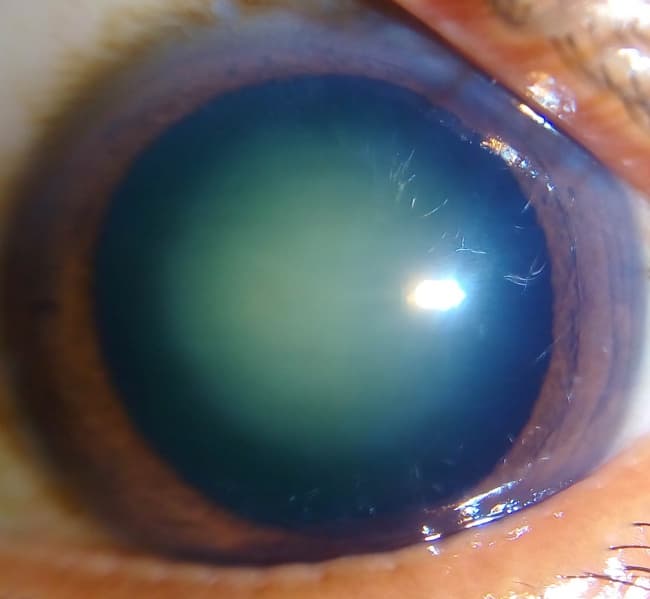
As we grow older our natural lenses become cloudy. This blocks the light from reaching the retina, resulting in blurred vision. Cataracts are completely treatable. The cloudy lenses are removed and replaced with an artificial lens that is implanted into the eye.
Cataracts can occur in adults and children due to numerous causes. The most common cause is age related changes. Other causes include diabetes, systemic diseases, and trauma.
Symptoms
Early symptoms include a decrease in vision. This can be either blurring of vision or loss of clarity. Other symptoms include hazy vision with glare and haloes at night.
Treatment
Cataract operations are the most common operations performed in medicine. It is usually done under local anaesthetic. The opacified lens is removed and an artificial lens is implanted. It has a extremely high success rate.
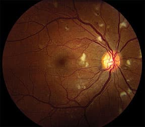
Diabetes is caused by high blood sugar levels and can affect many parts of the body. If the eyes are affected then it is called diabetic retinopathy. This is a potentially blinding condition. Therefore, patients with diabetes require regular eye examinations by an ophthalmologist.
The earlier diabetic eye disease is picked up the easier it is to treat. All patients with diabetes need to be screened at least on a yearly basis. The disease severity can vary from mild to profound, resulting in complete blindness. Diabetic eye disease can be divided into diabetic retinopathy (involving the retina) and diabetic maculopathy (swelling of the macula).
Treatment
There are different treatment options for the different stages of the disease. This varies from laser treatments of the retina, intravitreal injections, and surgery. Regular screening and early treatment plays an important role in the treatment of diabetic eye disease.
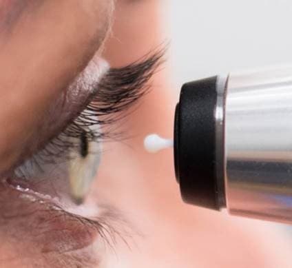
Glaucoma is caused by raised pressure inside the eye. This pressure causes damage to the optic nerve that exits the back of the eye. The diagnosis is made by examining the optic nerve and measuring the pressure in the eye.
Glaucoma is usually silent and only picked up when measuring the pressure or by examining the optic disc.
Treatment
It is treated with eye drops to decrease the pressure in the eye. If these drops do not work then laser treatment or surgery can be utilised.
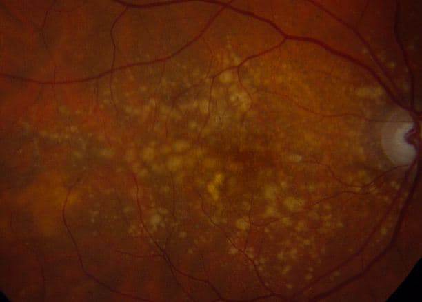
This is when the retinal pigment epithelial cells in the eye stop functioning optimally. There are two types of AMD: dry and wet. AMD is usually associated with old age and smoking.
Dry AMD is the most common form and involves degeneration of the nerve cells in the retina. It is usually a slow process with a permanent impact. Wet AMD occurs when the blood vessels in the retina leak fluid into the retina. This may cause a faster loss of vision.
Treatment
There is currently no cure for AMD. Dry AMD can be slowed down with vitamin supplementation. Wet AMD requires laser of the retina or intravitreal injections to prevent leakage from the blood vessels.
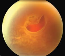
This is when the retinal tissue pulls away from the underlying supporting tissue. It usually starts due to a small hole or tear in the retina. This causes a localised detachment but can progress rapidly to a complete retinal detachment.
Symptoms
Symptoms usually include flashes of light with new floaters developing in the vision. This is followed by partial loss of vision and may progress to total loss of vision in the affected eye. It is more likely to develop in near-sighted individuals or if there is a history of trauma.
Treatment
Surgery is usually needed for retinal detachments. The outcome of the surgery will depend if the macula was involved in the detachment. Time is of the essence when treating retinal detachments as it can cause total loss of vision if left untreated.
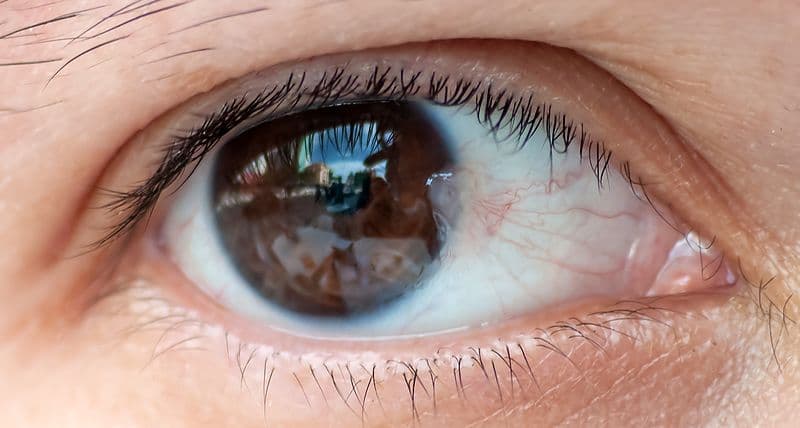
This is a common condition occurring in hot, dry areas. It is usually associated with sunlight exposure. It results in a small lump of tissue next to the cornea.
If left, this small lump of tissue can progress to form an unsightly mass that can eventually cause loss of vision. It may cause irritability and readiness of the eye.
Treatment
Pterygium is usually treated with surgical removal.
The Work Environment
Testimonials
Get in touch
Monday to Friday